
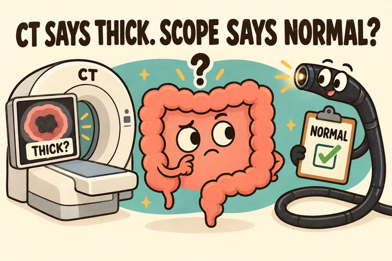
Colon wall thickening on a CT scan usually points to something real, but not always. Pool the studies, and about 3 in 4 people with it have a finding at colonoscopy, most often inflammation [1][2]. That leaves roughly 1 in 4 whose colonoscopy comes back normal [1][2]. It doesn’t mean either test lied. A squeezed or empty stretch of colon can look thick on CT, inflammation can heal before the scope gets there, and some problems sit outside the colon’s lining, where a scope can’t see.
If you read nothing else
- Your CT pictured the whole colon wall and everything around it, while your colonoscopy looked at the inside lining up close. Different jobs [4][5].
- A normal colon wall that’s squeezed down or empty can measure up to 5 mm on CT, versus 1 to 2 mm stretched open [6].
- Did your CT say “mass”? Take that seriously. In one study, all 12 people whose CT showed a mass had a precancerous polyp or a cancer at colonoscopy [10].
- Still having symptoms after a normal colonoscopy, check that the scope reached the end of the colon with a clean prep, then look beyond the colon [14][17].
The question, the way patients ask it
“I’m symptomatic, my CT scan shows that there’s something in my colon, but my colonoscopy is normal? How can that be?”
I get why that one drives people up the wall. Maybe yours started in the ER with belly pain, and somebody read you the words “colon wall thickening” off a report. You spent the next few weeks thinking the worst. Then you drank the prep (nobody has ever enjoyed that), and the colonoscopy came back clean. Two tests, two different stories, and a belly that still isn’t right.
Most of the time, though, both tests are telling you the truth. They’re looking at different things, and sometimes they’re looking on different days, which matters more than you’d think. Let’s sort it out.
CT scan vs colonoscopy: two different cameras
A CT scan (computed tomography, a ring-shaped X-ray machine that takes pictures in slices) shows your organs, soft tissue and blood vessels in a lot of detail [5]. One pass covers your colon, small bowel, appendix and, if you’re a woman, your uterus and ovaries [5]. In the colon, you get the whole thickness of the wall plus whatever’s sitting next to it.
A colonoscopy uses a flexible tube with a camera on the end [4]. It goes in through the rectum and drives up to where the small intestine opens into the colon. Then it comes back out slowly while your doctor examines the lining, the inside surface that faces the stool [4]. If something looks off, your doctor can take a biopsy, a small tissue sample, on the spot [4].
So the CT is the satellite view of the neighborhood, and the colonoscopy is me walking through the house with a flashlight. From above, a roof can look funny while every room is fine. And my flashlight can’t tell you a thing about the neighbor’s yard.
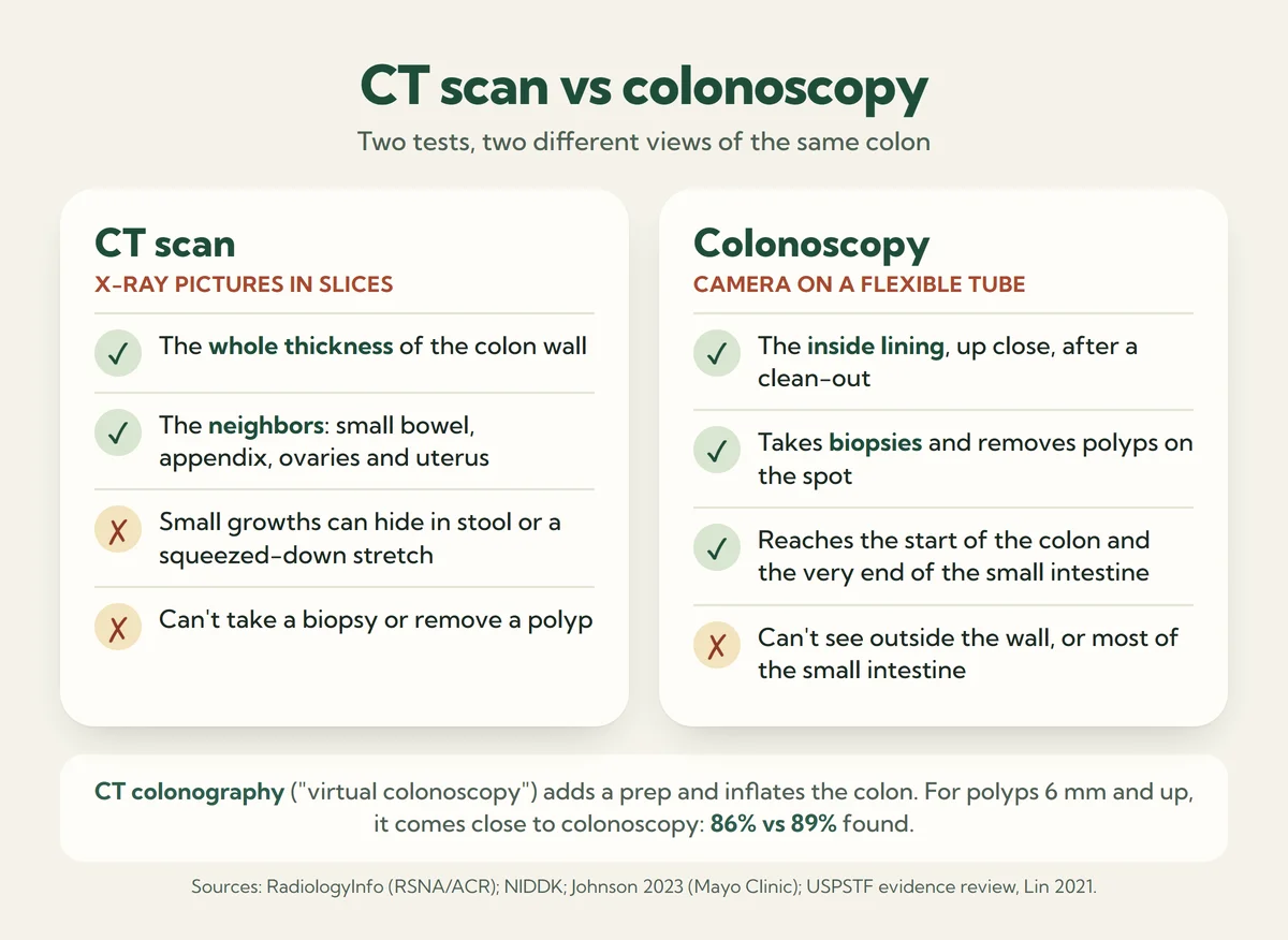
Heard of virtual colonoscopy? That’s CT colonography, a third test: you do a bowel prep, and your colon gets inflated with gas or air before the scan [8]. In screening studies it caught adenomas (the kind of polyp that can turn into cancer) 6 mm or larger nearly as often as colonoscopy, 86% versus 89% [9]. It’s a planned screening test, though, not the scan you get for belly pain.
Why a normal colon can look thick on CT
Your colon isn’t a pipe with a fixed wall. It squeezes, relaxes, and fills with gas and stool all day long, so how thick the wall looks depends on how stretched it is when the picture gets taken. On CT, a normal wall runs about 1 to 2 millimeters when it’s stretched open and up to 5 when it’s squeezed down or collapsed [6]. Same wall, same person, different moment.
A balloon does the same thing. Blown up, the rubber is thin. Let the air out and it goes thick and wrinkly, and nothing changed but the air.
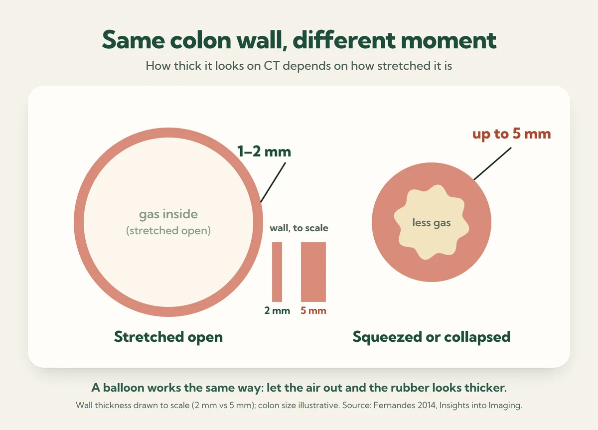
Radiologists know this trick well. One US study of colon wall thickening simply left out every scan where the colon wasn’t stretched open well enough to see clearly [2]. Does your report say “underdistended” or “collapsed”? That’s your radiologist flagging this exact problem [6].
Stool muddies things too. On CT colonography, stool can be hard to tell apart from a small polyp [8]. And when Mayo Clinic radiologists went back over the CT scans of people later diagnosed with colon cancer, the tumors they still couldn’t find were most likely small ones, hidden in colon contents and collapsed segments [7]. So a squeezed, half-full stretch of colon can fool a CT in either direction.
How often is colon wall thickening something real?
I won’t sugarcoat this part. Most of the time, thickening on CT does turn out to be something.
- Nine studies pooled, 1,252 people: about 3 in 4 had an abnormal colonoscopy [1]. When the CT looked like inflammation, the scope confirmed it 97% of the time [1].
- In Washington, DC (269 people), 27% had a normal colonoscopy. The rest had inflammation of the colon (32%), cancer (13%), colon ischemia (7%), inflammatory bowel disease (5%), or something else such as polyps or diverticulosis (16%) [2].
- Rhode Island, 107 people with belly pain: for 26%, nothing turned up that explained the CT. The most common finding was colon ischemia, a drop in blood flow to part of the colon, at 36%. Cancer was 7% [3].
- And in Brooklyn, 64% of 150 people whose emergency CT showed thickening had a matching finding when they were scoped [10].
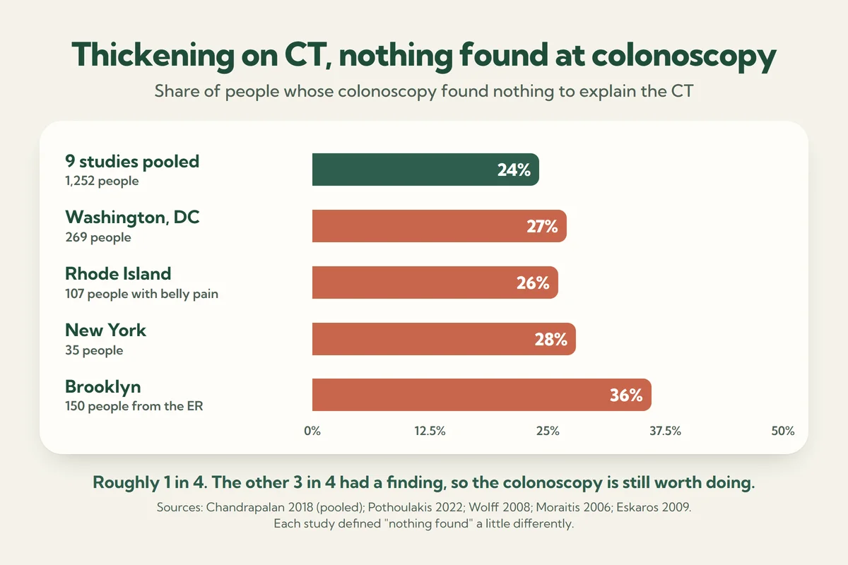
So a normal colonoscopy after CT thickening happens to roughly 1 in 4 people, give or take. That’s common. It’s also why the researchers behind these studies recommend the colonoscopy when a CT shows thickening [1][3][11].
One more number from Rhode Island stuck with me. Of the 18 people found to have cancer or inflammatory bowel disease, only 4 had bleeding or a low blood count [3]. No bleeding doesn’t mean nothing’s there.
When the CT says “mass”
Thickening and a mass are different words on a report. In the Brooklyn study, all 12 people whose CT showed a mass along with the thickening had an adenoma or a cancer at colonoscopy [10]. Pool the studies, and thickening that looked like cancer to the radiologist really was cancer 63% of the time [1]. So what if your report says mass and your scope says normal? Then everything rides on the quality of the colonoscopy. If it was a high-quality exam (it reached the end of the colon, the prep was clean, the lining was looked at carefully), I trust it: the mass isn’t there. If your symptoms come back later, I’d recommend either a repeat CT or a repeat colonoscopy.
Can a CT scan see colon cancer?
Often, yes, especially the bigger ones. But a regular CT isn’t built to hunt for colon cancer, and it misses some. Mayo Clinic radiologists studied 209 people with colon or rectal tumors who’d had a CT for some other reason in the year before diagnosis [7]:
- The original CT report mentioned the tumor in about half (51%).
- Looking back, knowing exactly where to look, they could spot another 32%.
- The last 18% stayed invisible even then. Those averaged 2.9 cm, about half the size of the tumors the reports caught [7].
So a CT can find colon cancer, and a normal CT doesn’t rule it out. For finding and removing polyps, colonoscopy is still the tool. Even when CT colonography finds a significant polyp, it comes out by colonoscopy [8].
A runner, a scary CT and a clean colonoscopy
A woman in her thirties came to see me a few weeks after a long-distance race. Late in the race she’d gotten sudden cramping, an urgent need to find a bathroom, and then bloody diarrhea. The ER did a CT. The report said thickening on the right side of her colon, and “neoplasm” (a growth) was on the list of possibilities. You can guess which word she remembered.
By the time I saw her, the bleeding had stopped within a couple of days and she felt pretty much normal. Her colonoscopy, three weeks after the race, was clean from end to end, and the biopsies were normal too. The most likely story was runner’s colon ischemia: blood flow to part of her colon dropped during the race, and it had already healed [12].
So who was right, the CT or the scope? Both of them. They just looked on different days.
Inflammation that healed before the scope got there
Timing matters more than most people realize. Colon ischemia (also called ischemic colitis) usually starts with sudden, mild cramping, an urgent need to go, and bright red or maroon blood within a day [12]. In more than half of cases it reverses on its own. Symptoms usually settle in 2 to 3 days, and the colon heals in 1 to 2 weeks [12]. Severe cases can take months [12].
The signs on colonoscopy fade just as fast. In one large study, the typical findings showed up in 47% of people scoped within 2 days, 33% at 2 to 5 days, and under 10% after day 5 [12]. That’s why the American College of Gastroenterology (ACG) recommends a colonoscopy within 48 hours when ischemia is suspected [12]. Book the scope three weeks out and there may be nothing left to see. Great news for your colon, even if it’s confusing for you.
Infections can thicken the wall too. In the Rhode Island study, 15% turned out to be infectious colitis [3].
When the CT is right and the scope can’t see it
A colonoscope works from the inside. Anything happening outside the lining, or beyond the scope’s reach, can show up on CT and still leave you with a perfectly normal colonoscopy.
- Diverticulitis, an infection or inflammation in small pouches that bulge out of the colon wall, is often diagnosed by CT [13]. Rarely, a cancer gets mistaken for diverticulitis. So if symptoms keep going after an attack, guidelines call for both imaging and a scope exam to sort it out [13].
- The appendix hangs off the start of the colon. Appendicitis was one of the causes behind colon thickening in the Rhode Island study [3].
- Your ovaries and uterus sit right next to the colon in the pelvis, and a CT shows them [5]. For some problems there, an MRI gives a better look [5].
- What about the small intestine? A colonoscopy can peek a short way into the ileum, the last part of it, and that’s about it [14]. The rest takes other tests, such as capsule endoscopy, a camera in a pill you swallow [14].
- Something pressing from the outside. The scope only sees the inside surface [4]. If a neighbor is leaning on the colon, the CT can show the bulge while the lining looks fine.
Then there’s one inside job a scope can miss by eye. With microscopic colitis, the lining usually looks completely normal, and only biopsies show the inflammation [15]. Its calling card is chronic, watery diarrhea without blood [16], so if that sounds like you, ask whether biopsies were taken.
Was your “normal” colonoscopy a complete one?
A normal report is only as good as the exam behind it. Check two things on yours:
- Did the scope reach the cecum, the pouch at the very start of the colon? Colonoscopists should get there in at least 95% of exams and photograph the landmarks to prove it [17]. If yours didn’t, part of your colon went unseen.
- How was the prep? Leftover stool can hide the lining [4]. Guidelines want an adequate prep in at least 90% of exams, and a poor prep usually means repeating the colonoscopy within a year [17]. If the CT spot was in a stretch the scope never saw cleanly and you’re still having symptoms, I’d want that look sooner.
And yes, even a complete, clean colonoscopy can miss something now and then. I go through how often, and what to ask your doctor, in Can a colonoscopy miss cancer?
Still having symptoms after a normal colonoscopy?
Symptoms that hang on deserve an answer, not a pat on the back. Here’s how I’d work through it with you:
- Get both reports in hand. Did the CT say thickening or mass, and where? Did the colonoscopy reach the cecum, with a good prep, and were biopsies taken [6][17]?
- Compare the pictures. I’ll pull up the CT images myself and line them up against what we saw during your colonoscopy.
- Look outside the colon. Where was the CT finding? Near the small bowel, that can mean a capsule endoscopy [14]. Near the ovaries or uterus, pelvic imaging [5]. After diverticulitis, imaging plus a scope exam [13].
- Diarrhea your main problem? Ask about biopsies [15].
- Redo what wasn’t complete. A poor prep or a scope that didn’t reach the cecum gets repeated [17].
If all of that comes back clean and you still hurt, conditions like IBS (irritable bowel syndrome, a gut that’s oversensitive without visible damage) are real and treatable. It’s not all in your head, either.
Family history counts too. If a parent, brother or sister had colon cancer or a large polyp, you may need colonoscopies earlier and more often. Here’s what a family history of colon polyps means for you.
When to see your doctor
Normal colonoscopy or not, don’t sit on these [18]:
- Blood in or on your stool
- Weight you’re losing without knowing why
- A low iron level in your blood (anemia)
- Belly pain, aches or cramps that don’t go away
- A change in your bowel habits, or feeling like you never quite empty
Frequently asked questions
What causes thickening of the colon wall?
Most often inflammation: an infection, colon ischemia (low blood flow), inflammatory bowel disease or diverticulitis [2][3]. Cancer is less common; it was 7% to 13% in two US studies [2][3]. And sometimes there’s nothing wrong at all, because a squeezed or empty colon can simply look thick on CT [6].
Is colon wall thickening cancer?
Can constipation or stool make the colon look thick on CT?
Can bowel wall thickening go away?
CT scan vs colonoscopy: which is more accurate?
Get the next one in your inbox
Join the Gutsavi newsletter for plain answers on gut health from a gastroenterologist, minus the jargon. Belly pain that won’t quit? Start at our abdominal pain and bloating hub. Seeing blood? Read Do I need a colonoscopy for rectal bleeding? There’s more in our Learn hub.
Sources
- Chandrapalan S, Tahir F, Kimani P, Sinha R, Arasaradnam R. Systematic review and meta-analysis: does colonic mural thickening on CT correlate with endoscopic findings at colonoscopy? Frontline Gastroenterol. 2018;9(4):278-84. doi:10.1136/flgastro-2018-100966
- Pothoulakis I, Wikholm C, Verma V, et al. Predictors of colorectal carcinoma and inflammatory bowel disease in patients with colonic wall thickening. JGH Open. 2022;6(3):159-65. PMC8938753
- Wolff JH, Rubin A, Potter JD, et al. Clinical significance of colonoscopic findings associated with colonic thickening on computed tomography: is colonoscopy warranted when thickening is detected? J Clin Gastroenterol. 2008;42(5):472-5. doi:10.1097/MCG.0b013e31804c7065
- National Institute of Diabetes and Digestive and Kidney Diseases (NIDDK). Colonoscopy. Reviewed August 2023. niddk.nih.gov
- RadiologyInfo.org (Radiological Society of North America and American College of Radiology). Computed tomography (CT): abdomen and pelvis. Reviewed June 2026. radiologyinfo.org
- Fernandes T, Oliveira MI, Castro R, et al. Bowel wall thickening at CT: simplifying the diagnosis. Insights Imaging. 2014;5(2):195-208. PMC3999365
- Johnson CD, Flicek KT, Mead-Harvey C, Quillen JK. Strategies for improving colorectal cancer detection with routine computed tomography. Abdom Radiol (NY). 2023;48(6):1891-9. PMC10036972
- RadiologyInfo.org (Radiological Society of North America and American College of Radiology). CT colonography or virtual colonoscopy. Reviewed June 2026. radiologyinfo.org
- Lin JS, Perdue LA, Henrikson NB, Bean SI, Blasi PR. Screening for colorectal cancer: updated evidence report and systematic review for the US Preventive Services Task Force. JAMA. 2021;325(19):1978-98. doi:10.1001/jama.2021.4417
- Eskaros S, Ghevariya V, Diamond I, Anand S. Correlation of incidental colorectal wall thickening at CT compared to colonoscopy. Emerg Radiol. 2009;16(6):473-6. doi:10.1007/s10140-009-0806-x
- Moraitis D, Singh P, Jayadevan R, Cayten CG. Colonic wall thickening on computed tomography scan and clinical correlation. Does it suggest the presence of an underlying neoplasia? Am Surg. 2006;72(3):269-71. PubMed 16553131
- Brandt LJ, Feuerstadt P, Longstreth GF, Boley SJ. ACG clinical guideline: epidemiology, risk factors, patterns of presentation, diagnosis, and management of colon ischemia (CI). Am J Gastroenterol. 2015;110(1):18-44. doi:10.1038/ajg.2014.395
- Peery AF, Shaukat A, Strate LL. AGA clinical practice update on medical management of colonic diverticulitis: expert review. Gastroenterology. 2021;160(3):906-11. doi:10.1053/j.gastro.2020.09.059
- NIDDK. Diagnosis of Crohn’s disease. Reviewed July 2024. niddk.nih.gov
- NIDDK. Diagnosis of microscopic colitis. Reviewed April 2021. niddk.nih.gov
- NIDDK. Symptoms & causes of microscopic colitis. Reviewed April 2021. niddk.nih.gov
- Rex DK, Anderson JC, Butterly LF, et al. Quality indicators for colonoscopy. Am J Gastroenterol. 2024;119(9):1754-80. doi:10.14309/ajg.0000000000002972
- Centers for Disease Control and Prevention (CDC). Symptoms of colorectal cancer. June 2026. cdc.gov
This article is for education only and isn’t medical advice. Talk with your own doctor about your care.
This article is for education only and isn't a substitute for advice from your own doctor. If something feels off, talk to your care team.